Digital Panoramic X-ray (OPG)
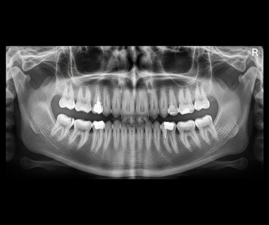
Digital Panoramic X-ray (OPG) The Digital Panoramic X-ray, also known as an Orthopantomogram (OPG), is a crucial diagnostic tool that provides a single, broad, two-dimensional view of the entire mouth. Unlike small intraoral X-rays that focus on only one or two teeth, the OPG captures the upper and lower jaws, all the teeth (erupted and unerupted), the surrounding bone, the nasal area, and the temporomandibular joints (TMJ). This comprehensive picture is indispensable for a foundational understanding of a patient’s overall oral health status. This imaging service is essential for initial comprehensive exams, orthodontic evaluations, and surgical planning. It is a fast, comfortable, and low-radiation way to screen for a wide array of conditions that cannot be detected during a visual clinical inspection, ensuring that our treatment plans are built on a complete and accurate understanding of your underlying anatomy. The Critical Broad Scope of OPG Imaging The OPG provides a snapshot of the entire oral landscape, allowing for the detection of critical issues that might be otherwise invisible: Assessment of Wisdom Teeth: It clearly visualizes the position, angulation, and stage of development of all four wisdom teeth, which is critical for planning preventative or surgical extractions. Jaw and Joint Health: It is the primary tool for evaluating the bone structure of the jaw (mandible and maxilla) and the condition of the temporomandibular joints (TMJs). Pathology Screening: It is used to screen for cysts, tumors, abscesses, and other bony lesions that develop silently within the jaw structure. Orthodontic and Implant Planning: It provides a clear view of tooth root length, the presence of missing or extra teeth, and bone height, offering foundational data for planning braces or dental implant placements. Periodontal Disease Assessment: It helps to assess the overall level of bone loss around the roots of all teeth, providing crucial insights into the severity and extent of periodontal disease. The Fast and Comfortable Digital Procedure The process of capturing a digital OPG is quick and non-invasive, requiring no placement of film inside the mouth: Positioning: The patient stands or sits with their chin resting on a small support, biting gently on a sterile bite-block to ensure the head is precisely aligned. The Scan: The machine's rotating arm moves in a semi-circle around the head. The entire exposure takes only a few seconds. Digital Advantages: The use of digital technology means significantly reduced radiation exposure compared to traditional film-based units. The image is instantly available on the computer screen for the doctor to review with the patient. Image Enhancement: Digital files allow for immediate manipulation of contrast, brightness, and magnification, enabling the dentist to perform advanced analysis without requiring a second image. Integrating OPG into Comprehensive Care The OPG often serves as the crucial first step in any comprehensive treatment journey. It flags potential issues—such as impacted teeth or early pathology—that then guide the need for more localized, detailed imaging like periapical X-rays or a focused CBCT scan. This systematic use of technology ensures we diagnose with accuracy and treat with confidence, prioritizing patient safety and predictive outcomes.