Cyst/Tumor Enucleation (Oral Surgery)
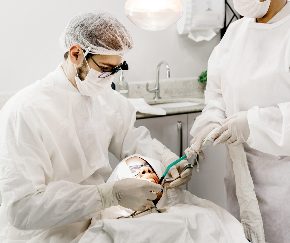
Cyst/Tumor Enucleation (Oral Surgery) The jawbones and surrounding soft tissues can occasionally develop fluid-filled sacs (cysts) or abnormal growths (benign tumors). While many of these are asymptomatic initially, they can cause significant problems if left untreated, including bone destruction, tooth displacement, and severe pain. Our Cyst/Tumor Enucleation service is a specialized oral surgery procedure dedicated to the complete and safe removal of these pathological lesions from the oral and maxillofacial region. The procedure of enucleation involves surgically removing the entire cyst or tumor mass without rupture, thereby maximizing the chances of complete healing and preventing recurrence. This surgery is often essential for preserving the integrity of the jawbone, protecting the vitality of adjacent teeth, and eliminating chronic sources of infection or potential malignancy. The Importance of Timely Surgical Removal Early diagnosis and intervention are critical when dealing with oral pathology. Jaw cysts and benign tumors pose several risks: Bone Destruction: As they grow, these lesions slowly resorb and replace healthy jawbone, severely weakening the structural support for teeth and implants. Tooth Mobility and Loss: Lesions near the root tips can compromise the periodontal ligament or push against the teeth, causing them to loosen, shift, or be displaced. Infection: Cysts can become secondarily infected, leading to acute abscesses, swelling, and systemic complications. Diagnostic Certainty: Enucleation allows for the complete removal and submission of the tissue to a pathologist. This definitive biopsy is crucial for determining the exact nature of the lesion (e.g., dentigerous cyst, ameloblastoma) and planning any necessary follow-up care. The Precision Surgical Enucleation Protocol Our surgical procedure is guided by advanced diagnostics to ensure maximal safety and minimal disruption to the surrounding healthy tissues: 3D Imaging (CBCT): We use CBCT Scans (Service ID 28) to precisely map the lesion's exact three-dimensional size, boundaries, and its relationship to vital structures like nerves, blood vessels, and the nasal or sinus cavities. Anesthesia and Exposure: The procedure is performed under local anesthesia with sedation options available. A small flap of gum tissue is gently lifted to expose the underlying jawbone where the lesion is located. Bone Access: A controlled, minimal amount of bone is removed to create an access window to the cyst or tumor capsule. Enucleation: The surgeon carefully separates the entire pathological sac from the surrounding bone wall, ensuring that the entire capsule is removed intact. Bone Regeneration and Suturing: The resulting bone cavity is thoroughly cleaned. In large defects, Bone Grafting (Service ID 16) material may be placed to encourage rapid bone healing and regeneration. The gum tissue is then sutured closed. Follow-Up and Histopathology The removed tissue is immediately sent for histopathological analysis. The results are critical for confirming the benign nature of the lesion or identifying any need for further treatment. We provide dedicated post-operative care instructions, monitor the healing process with follow-up appointments, and take periodic X-rays to confirm that the bone defect is successfully filling in with healthy, new bone tissue.