Microscopic Root Canal Treatment
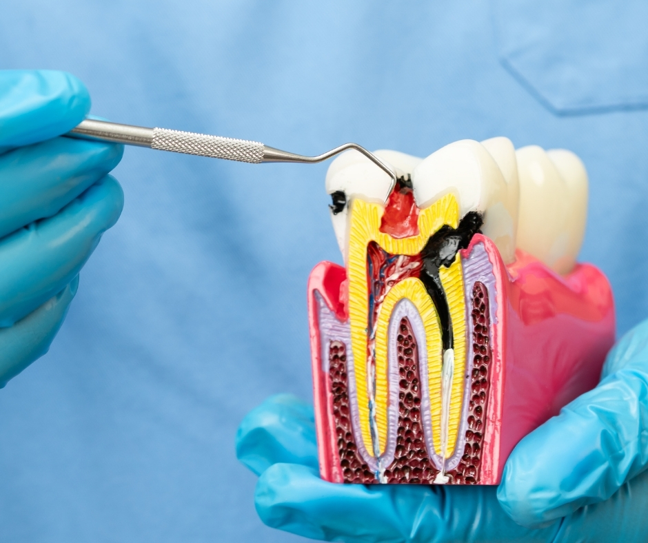
Microscopic Root Canal Treatment Root canal treatment is the process of saving a tooth that has a deeply infected or damaged nerve (pulp). While it can be done conventionally, our practice elevates this procedure by utilizing high-magnification dental operating microscopes. Microscopic Root Canal Treatment is the gold standard in endodontics, transforming a historically complex and sometimes unpredictable procedure into a routine, precise, and highly successful one. The microscope provides up to 25 times magnification and intense coaxial lighting, allowing the endodontist to see fine anatomical details that are invisible to the naked eye. This superior visualization is critical, as root canals are incredibly small, often curved, and contain complex lateral branches. By incorporating this technology, we maximize the chances of completely removing the infection while minimizing the removal of healthy tooth structure, preserving the long-term strength of your tooth. The Precision Advantage of the Microscope The use of the operating microscope significantly enhances every phase of the treatment, leading to better clinical outcomes: Locating Hidden Canals: The microscope allows us to locate tiny, calcified, or extra canals (which are often the reason for treatment failure) ensuring every source of infection is sealed. Fracture Detection: It enables the early identification of micro-fractures and cracks in the tooth and root, which may alter the prognosis and treatment plan. Foreign Body Retrieval: In re-treatment cases, the microscope is essential for locating and safely retrieving separated instruments or old filling materials from within the canal without damaging the root walls. Maximal Tooth Preservation: The enhanced vision permits the use of smaller, more conservative instruments, meaning we only remove what is infected and preserve the surrounding healthy dentin. The Treatment Process Our microscopic procedure follows a strict protocol to ensure safety and success: Anesthesia & Isolation: The tooth is made completely numb, and a protective rubber barrier (rubber dam) is placed to isolate the tooth from bacteria in the saliva. Access & Visualization: An access opening is made, and the operating microscope is positioned to illuminate and magnify the pulp chamber and canal entrances. Cleaning & Shaping: Using flexible, precision nickel-titanium rotary files under microscopic control, the canals are meticulously cleaned, disinfected, and shaped. Three-Dimensional Sealing: Once clean, the canal system is hermetically sealed with a thermoplastic filling material, ensuring a tight, three-dimensional seal from the top to the tip of the root. A Predictable and Comfortable Experience For the patient, the microscopic approach means more predictability and less time spent searching for canals. This efficiency reduces the overall time in the dental chair and significantly improves comfort. Furthermore, the high success rate of microscopic endodontics means you can have confidence that your natural tooth has been saved and is prepared to last for many years with a final protective crown.